🛑 NÜKLEİK ASİT
|
|
🛑 NÜKLEİK ASİT VE GEN ANLATIMI
NÜKLEİK ASİT VE GEN ANLATIMI
- Nükleik asitler doğal olarak oluşan kimyasal bileşimlerdir.
- Nükleik asitler hücrenin ana bilgi taşıyıcı molekülleridir ve protein bireşimini yöneterek her dirimlinin kalıtımsal özelliklerini belirlerler.
- Nükleik asitler kalıtımsal bilgiyi depolar, iletir, ve bu bilginin anlatımına katılır.
- İki tip nükleik asit, deoksinükleik asit (DNA) ve ribonükleik asit (RNA), dirimli örgenliklerin karmaşık bileşenlerini bir kuşaktan bir sonrakine yeniden üretmelerini sağlar.
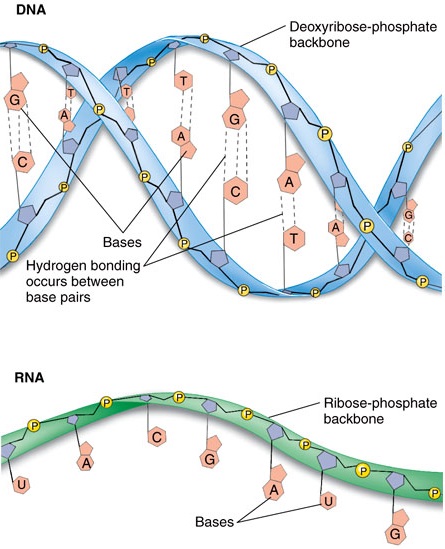
DNA ve RNA |
|
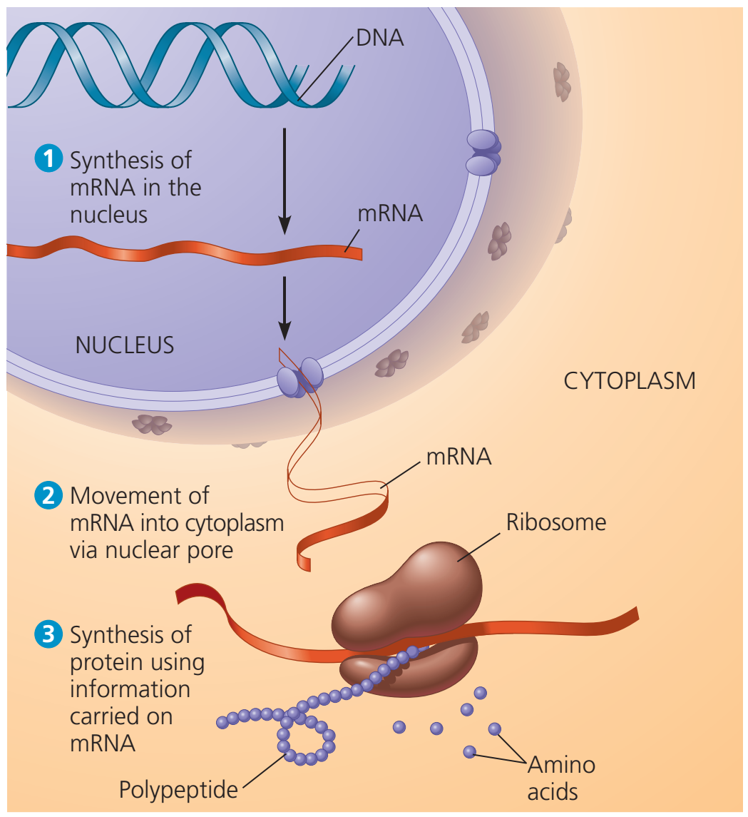
Gen Anlatımı: DNA → RNA → protein.
Bir ökaryotik hücrede, çekirdekteki DNA iletmen RNA (mRNA) bireşimini sağlayarak sitoplazmada protein üretimini programlar.
|
|
- DNA örgenliklerin ebeveynlerinden kalıt aldıkları genetik gereçtir.
- DNA kendi eşlenimi için yönergeler sağlar.
- DNA Gen Anlatımı denilen süreçte RNA bireşimini yönetir ve RNA yoluyla protein bireşimini denetler.
- Gen Anlatımı: DNA → RNA → protein.
- Bir ökaryotik hücrede, çekirdekteki DNA iletmen RNA (mRNA) bireşimini sağlayarak sitoplazmada protein üretimini programlar.
- Her bir kromozom genellikle yüzlerce ya da daha fazla gen taşıyan uzun bir DNA molekülünü kapsar.
- Bir hücre bölünerek kendini yeniden üretirken, DNA molekülleri eşlemlenir ve bir hücreler kuşağından bir sonrakine geçirilir.
- Hücrenin tüm etkinliklerini programlayan bilgiler DNAnın yapısında kodludur.
- Genetik gizilliği edimselleştirmek için, genotipi fenotipe çevirmek için proteinler gerekir.
- Bilgilerin DNAdan proteinlere akış süreci olan Gen Anlatımında öteki nükleik asit tipi olan RNA aracılık eder.
- DNA molekülü üzerinde verili bir gen iletmen RNA (mRNA) denilen RNA tipinin bireşimini yönetir.
- mRNA molekülü hücrenin protein-bireşimi yapan düzeneği (ribozom) ile etkileşim içinde bir polipeptidin üretimini yönetir; üretilen polipeptid sonra bir proteinin bir bölümünü ya da bütününü oluştumak üzere protein katlama sürecine girer.
- Protein bireşim siteleri ribozomlar denilen hücresel yapılardır.
- Bir ökaryotik hücrede ribozomlar sitoplazmada bulunur.
- mRNA proteinlerin yapımı için genetik bilgileri çekirdekten sitoplazmaya taşır.
- Prokaryotik hücrelerin çekirdekten yoksun olmalarına karşın, onlar da mRNA kullanır, ve bu molekül DNAnın bir iletisini amino asit dizilerine çeviren ribozomlara ve daha başka hücre donatımına taşır.
- Eşyazım sürecinde yeni bir tel oluşturmak üzere eşlemlenen DNA tel kalıp tel olarak adlandırılır.
- Kalıpta bilgiler saklanırken, ilk eşlemde tümleyici bir dizi vardır, özdeş değil.
- Eşlemin eşlemi kökensel diziyi bir kez daha üretir.
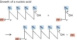
Bir nükleik asidin büyümesi. |
|
|
| |
|

|
|
|
|
🛑 NÜKLEİK ASİT YAPISI
NÜKLEİK ASİT YAPISI
- Nükleik asitler polinükleotid denilen polimerler olarak bulunan makromoleküllerdir.
- Her bir polinükleotid nükleotidler denilen monomerlerden oluşur.
- Bir nükleotid genel olarak üç parçadan oluşur: Bir beş-karbonlu şeker (bir pentoz); azot kapsayan bir baz (pürin ya da pirimidin); ve bir fosfat grubu.
- DNA adenin (A), guanin (G), sytozin (C), ve thymin (T) kapsar.
- RNA A, G, ve C kapsar, ama T yerine urasil (U) bulunur.
- DNAda deoksiriboz, RNAda riboz bulunur.
 or a deoxyribose sugar (in DNA).png)
Riboz ve deoksiriboz. |
|
- Polinükleotidler 3′, 5′-fosfodiester köprüleri ile bağlı nükleosidlerden oluşur.
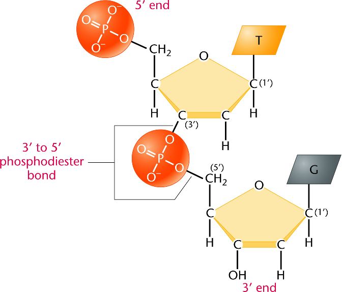
Fosfodiester bağ.
İki nükleotidin bir 3′—5′ fosfodiester bağ oluşumu yoluyla bir dinükleotid oluşturmak üzere bağlanması (şekerler deoksiribozdur). |
|
| |
| |
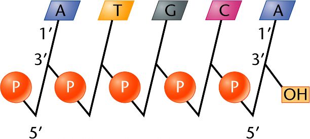
Çok sayıda fosfodiester bağ bir polinükleotid zincir oluşturur. |
|
- Genetik KOD polinükleotid zincir boyunca bazlar dizisinde bulunur.
- DNAda iki polinükleotid zincir bazları arasındaki eşleşme yoluyla birleştirilir (A T ve G C ile), ve bir çifte sarmal oluşturur.
- Bir zincir 5′ → 3′ yönünde, öteki 3′ → 5′ yönünde uzanır.
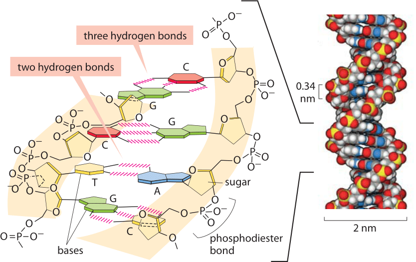
DNAda hidrojen ve fosfodiester bağları. |
|
- Bir polinükleotid üretmek için kullanılan başlangıç monomerinin üç fosfat grubu taşımasına karşın, polimerizasyon süreci sırasında ikisi yiter.
- Nükleotidin hiçbir fosfat grubu kapsamayan bölümüne nükleosid denir.
|
|
| |

|
|
|
|
🛑 NÜKLEİK ASİT ZİNCİR BÜYÜMESİ
NÜKLEİK ASİT ZİNCİRİNİN BÜYÜMESİ
- Hem DNA hem de RNA zincirleri kalıp DNA tellerinin eşlemlenmesi yoluyla üretilir.
- Nükleik asit zincirinin büyümesi nükleosidlerin 5′-trifosfatlarının eşlenim dizgesine eklenmesi ile sağlanır.
- Ekleme ribozun (deoksiribozun) 3′-OH ucunda yalnızca α fosfatın birleştirilmesi yoluyla yer alır (β ve γ fosfat grupları (pirofosfat) atılır).
- Nükleik asit zinciri ancak 3′-OH ucunda büyüyebilir.
- Zincirdeki ilk nükeotid tüm üç fosfatı tutabilir.
- Tek-telli DNA zinciri RNA zinciri ile aynı yolda uzar (ikisi arasındaki biricik ayrım DNAda 2′ deoksiriboz varken, RNAda aynı konumda OH bulunmasıdır.
|
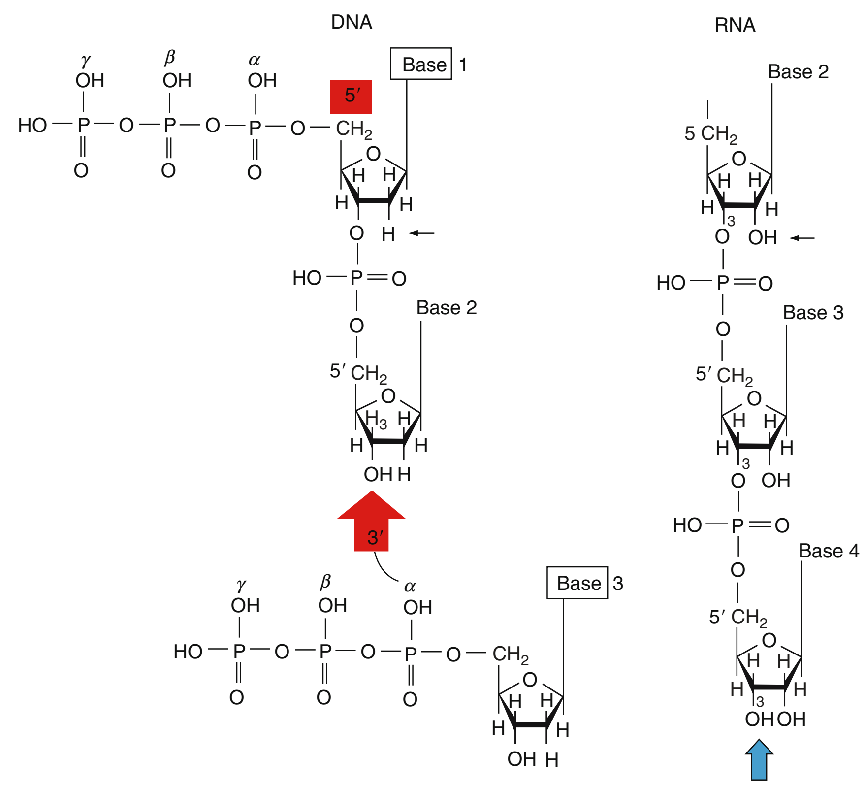
Nükleik asit zincirinin büyümesi.
|
📘 Nucleic Acid Chain Growth
Nucleic Acid Chain Growth
“Encyclopedia Of Genetics, Genomics, Proteomics And Informatics” / Springer (2008), p. 1372.
Nucleic Acid Chain Growth: Nucleic acid chain growth is secured by adding 5′-triphosphates of nucleosides to the replicating system. The additions take place at
the 3′-OH end of the ribose (deoxyribose) by joining
only the α phosphate; the β and γ phosphate groups
(pyrophosphate) are split off. Nucleic acid chains can
grow only at the 3′-OH terminus. The first nucleotide
in the chain may retain all three phosphates.
The single-stranded DNA chain elongates in the
same manner as the RNA strand. The only difference
between the two is that in the DNA (solid arrow)
there is 2′ deoxyribose, whereas in the RNA, at the
same position, there is OH.
|
|
|
|
|
|
| |

|
|
|
|
🛑 DNA BAZ ÇİFTLERİ
DNA BAZ ÇİFTLERİ
- Her bir azotlu bazın azot atomları kapsayan bir ya da iki halkası vardır (azotlu bazlar denmesinin nedeni azot atomlarının çözeltiden H+ ionları alarak bazlar gibi davranma eğiliminde olmasıdır).
DNA baz çiftleri. |
|
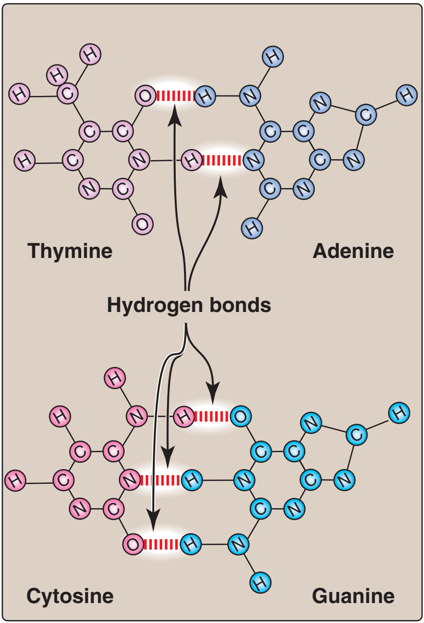
Tümleyici baz çiftleri arasındaki hidrojen bağları |
|
- İki baz ailesi vardır: Pirimidinler ve pürinler.
- Bir pirimidinin altı-üyeli bir karbon ve azot atomları halkası vardır.
- Pirimidin ailesinin üyeleri sitozin (C), thimin (T) ve urasildir (U).
- Pürinler adenin (A) ve guanindir (G).
- Pürinler bir beş-üyeli halkaya kaynaşmış altı-üyeli halka ile daha büyük moleküllerdir.
- Özgül pirimidinler ve pürinler halkalara bağlanan kimyasal gruplarında ayrım gösterir.
- Adenin, guanin ve sytozin hem DNAda hem de RNAda bulunur.
- Thimin yalnızca DNAda ve urasil yalnızca RNAda bulunur.
- DNAda şeker deoksiribozdur; RNAda riboz.
- Aralarındaki biricik ayrım deoksiribozun halkadaki ikinci karbon atomunda bir oksijen açığının olmasıdır ve deoksiriboz adı bundan doğar.
- Baz artı şeker bir nükleosid oluşturur.
- Bir nükleotidin yapısının tamamlanması için şekerin 5′ karbonuna bir ile üç arasında fosfat grubu eklenmelidir (şekerdeki karbon sayıları asallık simgesi olarak ′ kapsar.
- Bir fosfat eklendiğinde, bu molekül bir nükleosid monofosfattır; ama sık sık nükleotid olarak adlandırılır.
- Bir DNA (ya da mRNA) polimeri boyunca bazlar dizisi her bir gen için benzersizdir ve bu dizi hücreye özgü kalıtsal programı belirler.
- Genler yüzlerce ve binlerce nükleotid uzunluğunda olduğu için, olanaklı baz dizilerinin sayısı hemen hemen sınırsızdır.
- Gen tarafından taşınan bilgiler dört DNA bazının özgül dizilerinde kodlanır.
- Örneğin, 5′-AGGTAACTT-3′ dizisi belli bir anlama gelirken, 5′-CGCTTTAAC-3′ dizisinin başka bir anlamı vardır.
- Bir gendeki bazların doğrusal düzeni bir proteinin amino asit dizisini — birincil yapı — belirler ve bu da kendi payına proteinin hücredeki işlevini tanımlayan 3-D yapısını belirler.
- Ökaryotlarda DNA molekülleri histonlar ile etkileşime girerek nükleozom telleri oluşturur ve bunlar daha sıkı kıvrılı yapılara sarılır.
- Çekirdek içerisindeki kromozomlarda bulunan protein-DNA karmaşalarını anlatmak için kromatin terimi kullanılır.
- RNA tek-tellidir, ama teller geriye kendi üzerlerine döner ve baz çiftleri G—C ve A—U olarak birleşir.
- mRNAnın başı 5′ ucunda ve poli(A) kuyruğu 3′ ucundadır.
- tRNA birçok olağandışı nükleotid ve bir antikodon kapsayan bir yonca yaprağı yapısındadır.
|
| |
|
| |

|
|
|
|
🛑 DNA BAZ EŞLENMELERİ
DNA BAZ EŞLENMELERİ
- DNAdaki pürin ve pirimidin bazları hidrojen bağları ile bağlanır.
- A ve T değme durumunda iken iki hidrojen bağı oluşabilir; G ve C değme durumunda iken üç hidrojen bağı oluşabilir.
- Baz eşleşmesini hidrojen bağları belirler.
.png)
DNAda baz eşlenmesi. Nükleik asitlerde bazlar tümleyicidir. Her bir primidin yalnızca bir pürin baz ile eşlenir (G C ile, A T ile), ve bağ iki baz arasında oluşan hidrojen bağları yoluyla kurulur. |
|
- A T ile, ve G C ile, pürin pirimidin ile bağlanır (çift-telli molekülün genişliğinin göreli olarak değişmeden kalması bir pürinin bir primidin ile eşlenmesine bağlıdır).
- A’nın C ile değil de T ile eşlenmesinin nedeni T ile iki hidrojen bağı oluştururken C ile yalnızca bir hidrojen bağı oluşturabilmesidir.
- G C ile üç hidrojen bağı oluştururken, T ile yalnızca bir hidrojen bağı oluşturabilir.
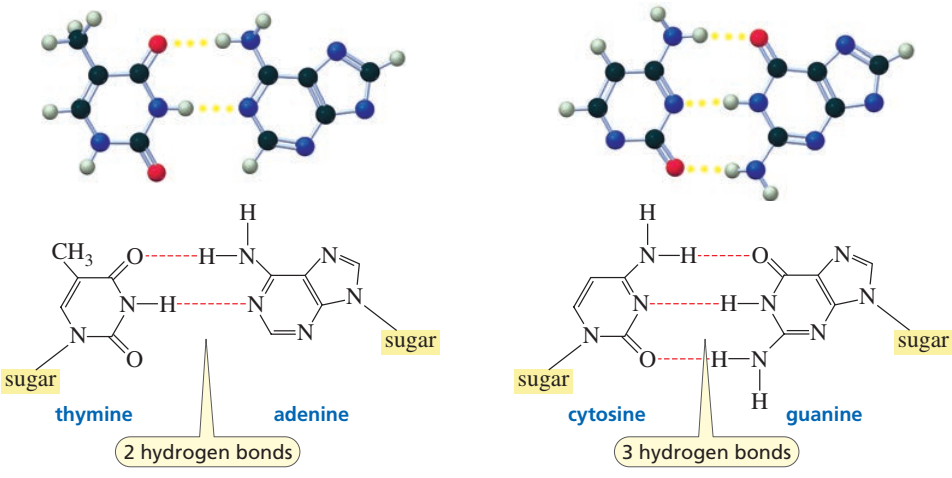
DNAda baz eşlenmesi: A ve T iki hidrojen bağı oluşturur; C ve G üç hidrojen bağı oluşturur. |
|
|
|
| |
|
|
|
🛑 NÜKLEOTİD
NÜKLEOTİD
- Nükleotid bir nükleosid ve bir fosfattan oluşur.
- DNAdaki her nükleotid aynı şekeri taşır, ama dört ayrı bazdan yalnızca birini taşır (A, C, G, T).
- Nükleotidler DNA ve RNA nükleik asit polimerlerinin monomerik birimleridir.
- Nükleotidler üç altbirimden oluşur: (1) bir azotlu baz; (2) beş-karbonlu şeker (riboz ya da deoksiriboz), ve bir ya da üç fosfattan oluşan bir fosfat grubu.
- Dört azotlu baz Guanin, Adenin, Sitozin ve Thimindir (RNAda thimin yerine Urasil geçer).
- Nükleotidler fosfatların sayısına göre (mono-, di-, tri-) ve şekerin tipine göre (deoksi- ya da yok), ve bazın tipine göre adlandırılır.
- Örnekler: ATP (adenozin trifosfat) adenin bazını, riboz şekerini, ve üç fosfat grubunu kapsar; dAMP bir adenin bazı, bir deoksiriboz şeker, ve bir fosfat grubu kapsar.
- Nükleotidler DNA ve RNA nükleik asit polimerlerinin monomerik birimleridir.
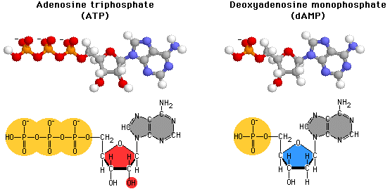
Dört azotlu baz Guanin, Adenin, Sitozin ve Thimindir (RNAda Thimin yerine Urasil geçer). |
|
- Ayrıca —
- Nükleotidler metabolizmada özsel bir rol oynar;
- Nükleosid trifosfatlar, adenozin trifosfat (ATP), guanosin trifosfat (GTP) sitidin trifosfat (CTP) ve üridin trifosfat (UTP) biçiminde, hücrenin çeşitli işlevleri için enerji sağlarlar;
- Hücre imleşimine katılırlar; ve
- Enzimatik tepkimelerde önemli kofaktörler olarak iş görürler.
|
|
| |

|
|
|
|
🛑 FOSFAT GRUBU
FOSFAT GRUBU
- Fosfat (PO43-) bir fosfat ve dört oksijen atomundan yapılı kimyasal bileşimdir.
- Fosfat karbon kapsayan bir moleküle bağlandığı zaman bileşime “fosfat grubu” adı verilir.
- Fosfat genetik gereçte bulunur.
- Fosfat adenozin trifosfatta (ATP) bulunur.
- Fosfat hücre membranını oluşturan fosfolipidlerde bulunur.
- Her bir nükleotidin 5-karbonlu şekeri ve fosfat grubu DNA ve RNAnın omurgasını oluşturmak üzere birleşir (nükleotidler nükleotidlere bağlanmadığı zaman, iki fosfat grubu daha alırlar).
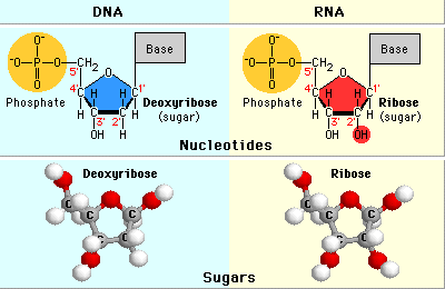
- Nükleotidler bir, iki ya da üç fosfat grubu kapsayabilir ve daha çok fosfat grubu daha çok enerji verimi demektir.
- Fosfat grupları fosfodiester bağları oluşturmak üzere birleşebilir. Fosfat grupları şeker gibi başka moleküller ile de birleşebilir.
- Fosfat nükleoside katıldığı zaman, moleküle nükleotid denir.
|
|
PROTEİNLERİN ETKİNLEŞTİRİLMESİ
- Fosfat grupları proteinleri hücrede tikel işlevlerini yerine getirebilmeleri için etkinleştirir.
- Proteinler bir fosfat grubunun eklenmesi demek olan fosforilasyon yoluyla etkinleştirilir.
- Protein fosforilasyonu tüm yaşam biçimlerinde yer alır.
- Defosforilasyon proteinlerin etkinliğini sonlandırır.
ENERJİ MOLEKÜLLERİNİN PARÇASI
- Adenezin trifosfat ya da ATP hücerelerdeki ana enerji kaynağıdır.
- ATP adenozin ve üç fosfattan yapılıdır.
- ATPden elde edilen enerji fosfatların kimyasal bağında taşınır.
- Fosfat bağlarının kırılması enerji üretimidir.
FOSFOLİPİDLERİN PARÇASI
- Fosfolipidler hücre membranının ve hücre çekirdeği kılıfının ana bileşenidir.
- Her fosfolipid bir lipid molekülünden ve bir fosfat grubundan yapılıdır.
- Fosfolipidler fosfolipid ikili-tabaka oluşturmak üzere sıralar olarak dizilir.
- Fosfolipid ikili-tabaka yarı-geçirgendir.
TAMPON İŞLEVİ
- Fosfat hücrelerde önemli bir tampondur ve bir özdeğin pHsını ne fazla asidik ne de fazla bazik olacak bir yolda yüksüz tutar.
BEDENDE
- İnsan bedendeki fosforun %85 kadarı kemiklerde ve dişlerde bulunur.
- Kalsiyum fosfat hem dişlere hem de kemiklere sert yapılarını verir.
- Fosfor bedende kalsiyumdan sonra en sık bulunan ikinci elementtir.
|
|
| |

|
|
|
|
🛑 BAZLAR VE GENETİK KOD
BAZLAR VE GENETİK KOD
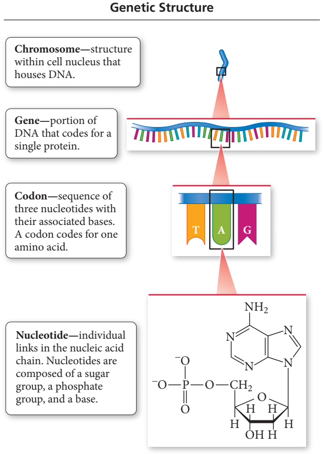
Genetik yapı. Genetik bilgilerin hiyerarşik yapısı kromozom, gen, kodon ve nükleotiddir. |
|
- Nükleik asit zincirindeki bazların sırası bir proteindeki amino asitlerin dizisini belirler.
- Yalnızca 4 baz vardır.
- 4 baz 20 amino asidi kodlamalıdır.
- 20 çeşit amino asit ve 4 çeşit baz olduğuna göre, tek bir baz tek bir amino asit için kodlama yapamaz
- Her bir amino asit için üç nükleotidden oluşan bir dizi kodlama yapar.
- Bir amino asit için kodlama yapan üçlü nükleotid dizisine kodon denir.
- Her bir tikel kodon tarafından belirlenen amino asidi tanımlayan Genetik Kod 1961’de geliştirildi.
- Hemen hemen tüm örgenliklerde aynı kodonlar aynı amino asidi kodlar.
- Örneğin bakteride ya da insanda DNAda AGT dizisi serin amino asidini kodlar; ACC dizisi thereonin amino asidini kodlar.
- Bir gen DNA molekülünde tek bir proteini kodlayan bir kodonlar dizisidir.
- Proteinler büyüklükte birkaç düzine amiono asitten binlerce amino aside değiştiği için, genler büyüklükte düzinelerden binlerce kodona değişir.
- Örneğin yumurta-akı lisozimi 129 amino asitten oluşur ve buna göre lisozim geni 129 kodon kapsar.
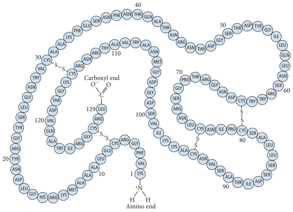
Yumurta-akı Lisozimin birincil yapısı. Bir proteinde birincil yapı amino asitlerin dizisini belirtir. |
|
- Her bir kodon tek bir amino asidi belirleyen üç harfli bir sözcük gibidir.
- Genler kromozomlar denilen yapılarda kapsanır.
|
|
| |

|
|
|
|
| |

|
|
|
|
Nucleic acid (B)
Nucleic acid (B)
Introduction
|
Introduction
Introduction (B)
Nucleic acid, naturally occurring chemical compound that is capable of being broken down to yield phosphoric acid, sugars, and a mixture of organic bases (purines and pyrimidines).
Nucleic acids are the main information-carrying molecules of the cell, and, by directing the process of protein synthesis, they determine the inherited characteristics of every living thing.
The two main classes of nucleic acids are deoxyribonucleic acid (DNA) and ribonucleic acid (RNA).
DNA is the master blueprint for life and constitutes the genetic material in all free-living organisms and most viruses.
RNA is the genetic material of certain viruses, but it is also found in all living cells, where it plays an important role in certain processes such as the making of proteins. |
| |
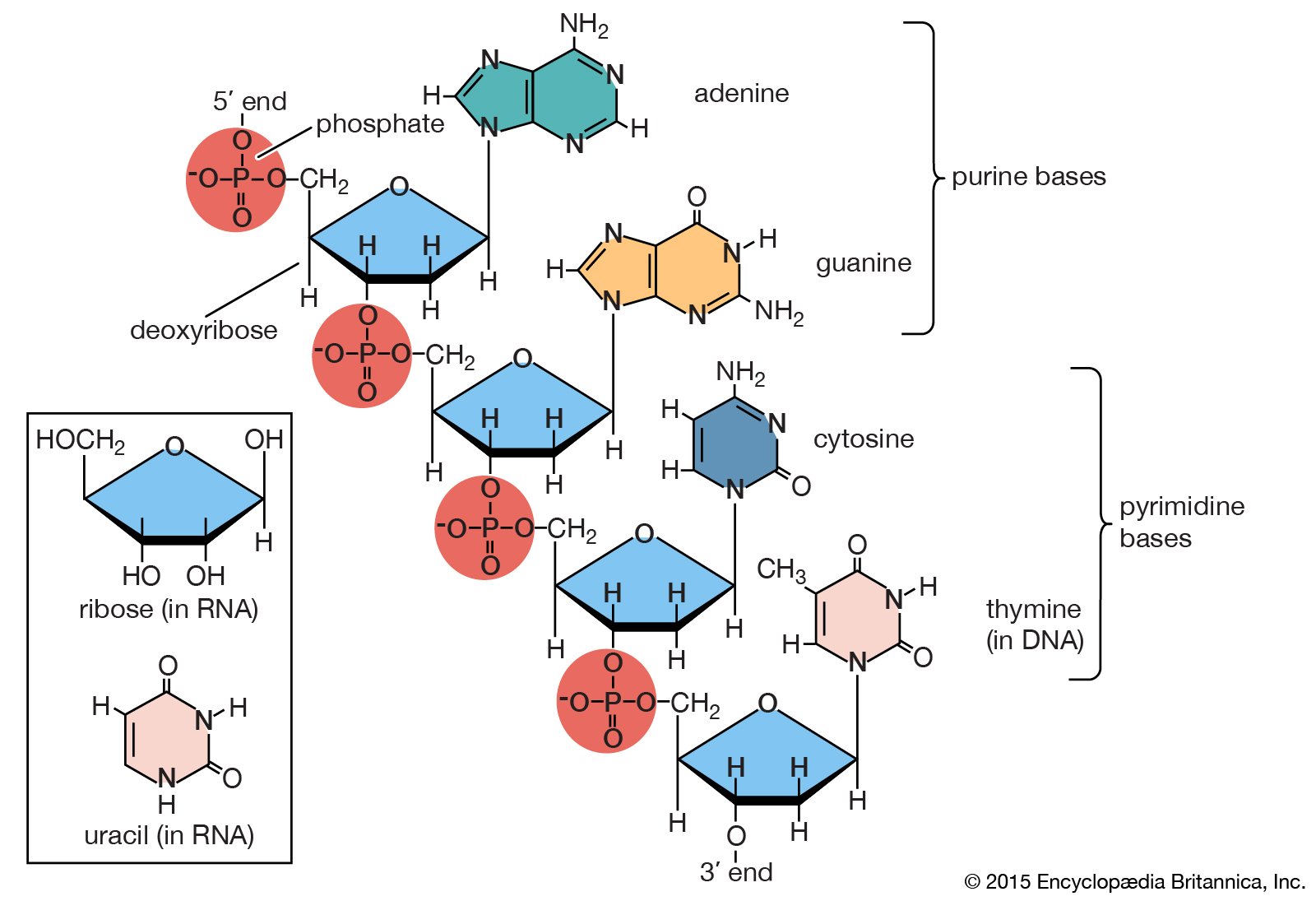
Polynucleotide chain of deoxyribonucleic acid (DNA)
Portion of polynucleotide chain of deoxyribonucleic acid (DNA). The inset shows the corresponding pentose sugar and pyrimidine base in ribonucleic acid (RNA).
. |
|
|
|
| |
| This article covers the chemistry of nucleic acids, describing the structures and properties that allow them to serve as the transmitters of genetic information. For a discussion of the genetic code, see heredity, and for a discussion of the role played by nucleic acids in protein synthesis, see metabolism. |

|
|
|
|
| |
Nucleotides: Building Blocks Of Nucleic Acids
|
Basic structure
Basic structure (B)
Nucleic acids are polynucleotides — that is, long chainlike molecules composed of a series of nearly identical building blocks called nucleotides. Each nucleotide consists of a nitrogen-containing aromatic base attached to a pentose (five-carbon) sugar, which is in turn attached to a phosphate group. Each nucleic acid contains four of five possible nitrogen-containing bases: adenine (A), guanine (G), cytosine (C), thymine (T), and uracil (U). A and G are categorized as purines, and C, T, and U are collectively called pyrimidines.
All nucleic acids contain the bases A, C, and G; T, however, is found only in DNA, while U is found in RNA.
The pentose sugar in DNA (2′-deoxyribose) differs from the sugar in RNA (ribose) by the absence of a hydroxyl group (―OH) on the 2′ carbon of the sugar ring.
Without an attached phosphate group, the sugar attached to one of the bases is known as a nucleoside.
The phosphate group connects successive sugar residues by bridging the 5′-hydroxyl group on one sugar to the 3′-hydroxyl group of the next sugar in the chain.
These nucleoside linkages are called phosphodiester bonds and are the same in RNA and DNA. |

|
|
|
|
Biosynthesis and degradation
Biosynthesis and degradation (B)
Nucleotides are synthesized from readily available precursors in the cell. The ribose phosphate portion of both purine and pyrimidine nucleotides is synthesized from glucose via the pentose phosphate pathway. The six-atom pyrimidine ring is synthesized first and subsequently attached to the ribose phosphate. The two rings in purines are synthesized while attached to the ribose phosphate during the assembly of adenine or guanine nucleosides. In both cases the end product is a nucleotide carrying a phosphate attached to the 5′ carbon on the sugar. Finally, a specialized enzyme called a kinase adds two phosphate groups using adenosine triphosphate (ATP) as the phosphate donor to form ribonucleoside triphosphate, the immediate precursor of RNA. For DNA, the 2′-hydroxyl group is removed from the ribonucleoside diphosphate to give deoxyribonucleoside diphosphate. An additional phosphate group from ATP is then added by another kinase to form a deoxyribonucleoside triphosphate, the immediate precursor of DNA.
During normal cell metabolism, RNA is constantly being made and broken down. The purine and pyrimidine residues are reused by several salvage pathways to make more genetic material. Purine is salvaged in the form of the corresponding nucleotide, whereas pyrimidine is salvaged as the nucleoside. |

|
|
|
|
| |
Deoxyribonucleic Acid (DNA)
|
Deoxyribonucleic Acid (DNA)
Deoxyribonucleic Acid (DNA) (B)
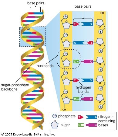
DNA is a polymer of the four nucleotides A, C, G, and T, which are joined through a backbone of alternating phosphate and deoxyribose sugar residues. These nitrogen-containing bases occur in complementary pairs as determined by their ability to form hydrogen bonds between them.
A always pairs with T through two hydrogen bonds, and G always pairs with C through three hydrogen bonds. The spans of A:T and G:C hydrogen-bonded pairs are nearly identical, allowing them to bridge the sugar-phosphate chains uniformly. This structure, along with the molecule’s chemical stability, makes DNA the ideal genetic material. The bonding between complementary bases also provides a mechanism for the replication of DNA and the transmission of genetic information. |
| |
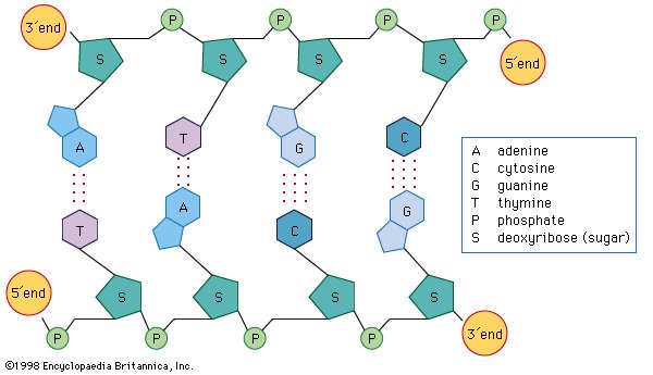
DNA structure, showing the nucleotide bases cytosine (C), thymine (T), adenine (A), and guanine (G) linked to a backbone of alternating phosphate (P) and deoxyribose sugar (S) groups. Two sugar-phosphate chains are paired through hydrogen bonds between A and T and between G and C, thus forming the twin-stranded double helix of the DNA molecule. |
|
|
|

|
|
|
|
Chemical structure
Chemical structure (B)
Chemical structure
In 1953 James D. Watson and Francis H.C. Crick proposed a three-dimensional structure for DNA based on low-resolution X-ray crystallographic data and on Erwin Chargaff’s observation that, in naturally occurring DNA, the amount of T equals the amount of A and the amount of G equals the amount of C. Watson and Crick, who shared a Nobel Prize in 1962 for their efforts, postulated that two strands of polynucleotides coil around each other, forming a double helix. The two strands, though identical, run in opposite directions as determined by the orientation of the 5′ to 3′ phosphodiester bond. The sugar-phosphate chains run along the outside of the helix, and the bases lie on the inside, where they are linked to complementary bases on the other strand through hydrogen bonds. |
| |
The initial proposal of the structure of DNA by James Watson and Francis Crick, which was accompanied by a suggestion on the means of replication. |
|
|
|
| |
The double helical structure of normal DNA takes a right-handed form called the B-helix. The helix makes one complete turn approximately every 10 base pairs. B-DNA has two principal grooves, a wide major groove and a narrow minor groove. Many proteins interact in the space of the major groove, where they make sequence-specific contacts with the bases. In addition, a few proteins are known to make contacts via the minor groove.
Several structural variants of DNA are known. In A-DNA, which forms under conditions of high salt concentration and minimal water, the base pairs are tilted and displaced toward the minor groove. Left-handed Z-DNA forms most readily in strands that contain sequences with alternating purines and pyrimidines. DNA can form triple helices when two strands containing runs of pyrimidines interact with a third strand containing a run of purines.
B-DNA is generally depicted as a smooth helix; however, specific sequences of bases can distort the otherwise regular structure. For example, short tracts of A residues interspersed with short sections of general sequence result in a bent DNA molecule. Inverted base sequences, on the other hand, produce cruciform structures with four-way junctions that are similar to recombination intermediates. Most of these alternative DNA structures have only been characterized in the laboratory, and their cellular significance is unknown. |

|
|
|
|
Biological structures
Biological structures (B)
Naturally occurring DNA molecules can be circular or linear. The genomes of single-celled bacteria and archaea (the prokaryotes), as well as the genomes of mitochondria and chloroplasts (certain functional structures within the cell), are circular molecules. In addition, some bacteria and archaea have smaller circular DNA molecules called plasmids that typically contain only a few genes. Many plasmids are readily transmitted from one cell to another. For a typical bacterium, the genome that encodes all of the genes of the organism is a single contiguous circular molecule that contains a half million to five million base pairs. The genomes of most eukaryotes and some prokaryotes contain linear DNA molecules called chromosomes. Human DNA, for example, consists of 23 pairs of linear chromosomes containing three billion base pairs.
In all cells, DNA does not exist free in solution but rather as a protein-coated complex called chromatin. In prokaryotes, the loose coat of proteins on the DNA helps to shield the negative charge of the phosphodiester backbone. Chromatin also contains proteins that control gene expression and determine the characteristic shapes of chromosomes. In eukaryotes, a section of DNA between 140 and 200 base pairs long winds around a discrete set of eight positively charged proteins called a histone, forming a spherical structure called the nucleosome. Additional histones are wrapped by successive sections of DNA, forming a series of nucleosomes like beads on a string. Transcription and replication of DNA is more complicated in eukaryotes because the nucleosome complexes have to be at least partially disassembled for the processes to proceed effectively. |
| |
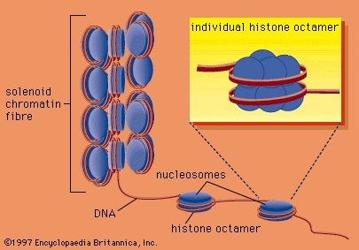
DNA wrapped around clusters of histone proteins to form nucleosomes, which are coiled to form solenoids, the basis of the chromatin fibre that makes up chromosomes. |
|
|
|
| |
| Most prokaryote viruses contain linear genomes that typically are much shorter and contain only the genes necessary for viral propagation. Bacterial viruses called bacteriophages (or phages) may contain both linear and circular forms of DNA. For instance, the genome of bacteriophage λ (lambda), which infects the bacterium Escherichia coli, contains 48,502 base pairs and can exist as a linear molecule packaged in a protein coat. The DNA of phage λ can also exist in a circular form (as described in the section Site-specific recombination) that is able to integrate into the circular genome of the host bacterial cell. Both circular and linear genomes are found among eukaryotic viruses, but they more commonly use RNA as the genetic material. |

|
|
|
|
Biochemical properties
Biochemical properties (B)
No text. |
|
|
|
Denaturation
Denaturation (B)
The strands of the DNA double helix are held together by hydrogen bonding interactions between the complementary base pairs. Heating DNA in solution easily breaks these hydrogen bonds, allowing the two strands to separate—a process called denaturation or melting. The two strands may reassociate when the solution cools, reforming the starting DNA duplex—a process called renaturation or hybridization. These processes form the basis of many important techniques for manipulating DNA. For example, a short piece of DNA called an oligonucleotide can be used to test whether a very long DNA sequence has the complementary sequence of the oligonucleotide embedded within it. Using hybridization, a single-stranded DNA molecule can capture complementary sequences from any source. Single strands from RNA can also reassociate. DNA and RNA single strands can form hybrid molecules that are even more stable than double-stranded DNA. These molecules form the basis of a technique that is used to purify and characterize messenger RNA (mRNA) molecules corresponding to single genes. |

|
|
|
|
Ultraviolet absorption
Ultraviolet absorption (B)
DNA melting and reassociation can be monitored by measuring the absorption of ultraviolet (UV) light at a wavelength of 260 nanometres (billionths of a metre). When DNA is in a double-stranded conformation, absorption is fairly weak, but when DNA is single-stranded, the unstacking of the bases leads to an enhancement of absorption called hyperchromicity. Therefore, the extent to which DNA is single-stranded or double-stranded can be determined by monitoring UV absorption. |
|
|
|
Chemical modification
Chemical modification (B)
After a DNA molecule has been assembled, it may be chemically modified—sometimes deliberately by special enzymes called DNA methyltransferases and sometimes accidentally by oxidation, ionizing radiation, or the action of chemical carcinogens. DNA can also be cleaved and degraded by enzymes called nucleases. |
|
|
|
Methylation
Methylation (B)
Three types of natural methylation have been reported in DNA. Cytosine can be modified either on the ring to form 5-methylcytosine or on the exocyclic amino group to form N4-methylcytosine. Adenine may be modified to form N6-methyladenine. N4-methylcytosine and N6-methyladenine are found only in bacteria and archaea, whereas 5-methylcytosine is widely distributed. Special enzymes called DNA methyltransferases are responsible for this methylation; they recognize specific sequences within the DNA molecule so that only a subset of the bases is modified. Other methylations of the bases or of the deoxyribose are sometimes induced by carcinogens. These usually lead to mispairing of the bases during replication and have to be removed if they are not to become mutagenic. |
|
|
|
Nucleases
Nucleases (B)
Nucleases are enzymes that hydrolytically cleave the phosphodiester backbone of DNA. Endonucleases cleave in the middle of chains, while exonucleases operate selectively by degrading from the end of the chain. Nucleases that act on both single- and double-stranded DNA are known.
Restriction endonucleases are a special class that recognize and cleave specific sequences in DNA. Type II restriction endonucleases always cleave at or near their recognition sites. They produce small, well-defined fragments of DNA that help to characterize genes and genomes and that produce recombinant DNAs. Fragments of DNA produced by restriction endonucleases can be moved from one organism to another. In this way it has been possible to express proteins such as human insulin in bacteria. |
|
|
|
Mutation
Mutation (B)
Chemical modification of DNA can lead to mutations in the genetic material. Anions such as bisulfite can deaminate cytosine to form uracil, changing the genetic message by causing C-to-T transitions. Exposure to acid causes the loss of purine residues, though specific enzymes exist in cells to repair these lesions. Exposure to UV light can cause adjacent pyrimidines to dimerize, while oxidative damage from free radicals or strong oxidizing agents can cause a variety of lesions that are mutagenic if not repaired. Halogens such as chlorine and bromine react directly with uracil, adenine, and guanine, giving substituted bases that are often mutagenic. Similarly, nitrous acid reacts with primary amine groups—for example, converting adenosine into inosine—which then leads to changes in base pairing and mutation. Many chemical mutagens, such as chlorinated hydrocarbons and nitrites, owe their toxicity to the production of halides and nitrous acid during their metabolism in the body. |

|
|
|
|
Supercoiling
Supercoiling (B)
Circular DNA molecules such as those found in plasmids or bacterial chromosomes can adopt many different topologies. One is active supercoiling, which involves the cleavage of one DNA strand, its winding one or more turns around the complementary strand, and then the resealing of the molecule. Each complete rotation leads to the introduction of one supercoiled turn in the DNA, a process that can continue until the DNA is fully wound and collapses on itself in a tight ball. Reversal is also possible. Special enzymes called gyrases and topoisomerases catalyze the winding and relaxation of supercoiled DNA. In the linear chromosomes of eukaryotes, the DNA is usually tightly constrained at various points by proteins, allowing the intervening stretches to be supercoiled. This property is partially responsible for the great compaction of DNA that is necessary to fit it within the confines of the cell. The DNA in one human cell would have an extended length of between two and three metres, but it is packed very tightly so that it can fit within a human cell nucleus that is 10 micrometres in diameter. |

|
|
|
|
Sequence determination
Sequence determination (B)
Methods to determine the sequences of bases in DNA were pioneered in the 1970s by Frederick Sanger and Walter Gilbert, whose efforts won them a Nobel Prize in 1980. The Gilbert-Maxam method relies on the different chemical reactivities of the bases, while the Sanger method is based on enzymatic synthesis of DNA in vitro. Both methods measure the distance from a fixed point on DNA to each occurrence of a particular base—A, C, G, or T. DNA fragments obtained from a series of reactions are separated according to length in four “lanes” by gel electrophoresis. Each lane corresponds to a unique base, and the sequence is read directly from the gel. The Sanger method has now been automated using fluorescent dyes to label the DNA, and a single machine can produce tens of thousands of DNA base sequences in a single run. |
|
|
|
| |
Ribonucleic Acid (RNA)
|
Ribonucleic Acid (RNA)
Ribonucleic Acid (RNA) (B)
RNA is a single-stranded nucleic acid polymer of the four nucleotides A, C, G, and U joined through a backbone of alternating phosphate and ribose sugar residues. It is the first intermediate in converting the information from DNA into proteins essential for the working of a cell. Some RNAs also serve direct roles in cellular metabolism. RNA is made by copying the base sequence of a section of double-stranded DNA, called a gene, into a piece of single-stranded nucleic acid. This process, called transcription (see below RNA metabolism), is catalyzed by an enzyme called RNA polymerase. |
|
|
|
Chemical structure
Chemical structure (B)
Whereas DNA provides the genetic information for the cell and is inherently quite stable, RNA has many roles and is much more reactive chemically. RNA is sensitive to oxidizing agents such as periodate that lead to opening of the 3′-terminal ribose ring. The 2′-hydroxyl group on the ribose ring is a major cause of instability in RNA, because the presence of alkali leads to rapid cleavage of the phosphodiester bond linking ribose and phosphate groups. In general, this instability is not a significant problem for the cell, because RNA is constantly being synthesized and degraded.
Interactions between the nitrogen-containing bases differ in DNA and RNA. In DNA, which is usually double-stranded, the bases in one strand pair with complementary bases in a second DNA strand. In RNA, which is usually single-stranded, the bases pair with other bases within the same molecule, leading to complex three-dimensional structures. Occasionally, intermolecular RNA/RNA duplexes do form, but they form a right-handed A-type helix rather than the B-type DNA helix. Depending on the amount of salt present, either 11 or 12 base pairs are found in each turn of the helix. Helices between RNA and DNA molecules also form; these adopt the A-type conformation and are more stable than either RNA/RNA or DNA/DNA duplexes. Such hybrid duplexes are important species in biology, being formed when RNA polymerase transcribes DNA into mRNA for protein synthesis and when reverse transcriptase copies a viral RNA genome such as that of the human immunodeficiency virus (HIV).
Single-stranded RNAs are flexible molecules that form a variety of structures through internal base pairing and additional non-base pair interactions. They can form hairpin loops such as those found in transfer RNA (tRNA), as well as longer-range interactions involving both the bases and the phosphate residues of two or more nucleotides. This leads to compact three-dimensional structures. Most of these structures have been inferred from biochemical data, since few crystallographic images are available for RNA molecules. In some types of RNA, a large number of bases are modified after the RNA is transcribed. More than 90 different modifications have been documented, including extensive methylations and a wide variety of substitutions around the ring. In some cases these modifications are known to affect structure and are essential for function. |

|
|
|
|
Types of RNA
Types of RNA (B)
No text. |
|
|
|
Messenger RNA (mRNA
Messenger RNA (mRNA (B)
Messenger RNA (mRNA) delivers the information encoded in one or more genes from the DNA to the ribosome, a specialized structure, or organelle, where that information is decoded into a protein. In prokaryotes, mRNAs contain an exact transcribed copy of the original DNA sequence with a terminal 5′-triphosphate group and a 3′-hydroxyl residue. In eukaryotes the mRNA molecules are more elaborate. The 5′-triphosphate residue is further esterified, forming a structure called a cap. At the 3′ ends, eukaryotic mRNAs typically contain long runs of adenosine residues (polyA) that are not encoded in the DNA but are added enzymatically after transcription. Eukaryotic mRNA molecules are usually composed of small segments of the original gene and are generated by a process of cleavage and rejoining from an original precursor RNA (pre-mRNA) molecule, which is an exact copy of the gene (as described in the section Splicing). In general, prokaryotic mRNAs are degraded very rapidly, whereas the cap structure and the polyA tail of eukaryotic mRNAs greatly enhance their stability. |

|
|
|
|
Ribosomal RNA (rRNA)
Ribosomal RNA (rRNA) (B)
Ribosomal RNA (rRNA) molecules are the structural components of the ribosome. The rRNAs form extensive secondary structures and play an active role in recognizing conserved portions of mRNAs and tRNAs. They also assist with the catalysis of protein synthesis. In the prokaryote E. coli, seven copies of the rRNA genes synthesize about 15,000 ribosomes per cell. In eukaryotes the numbers are much larger. Anywhere from 50 to 5,000 sets of rRNA genes and as many as 10 million ribosomes may be present in a single cell. In eukaryotes these rRNA genes are looped out of the main chromosomal fibres and coalesce in the presence of proteins to form an organelle called the nucleolus. The nucleolus is where the rRNA genes are transcribed and the early assembly of ribosomes takes place.
|
|
|
|
Transfer RNA (tRNA)
Transfer RNA (tRNA) (B)
Transfer RNA (tRNA) carries individual amino acids into the ribosome for assembly into the growing polypeptide chain. The tRNA molecules contain 70 to 80 nucleotides and fold into a characteristic cloverleaf structure. Specialized tRNAs exist for each of the 20 amino acids needed for protein synthesis, and in many cases more than one tRNA for each amino acid is present. The nucleotide sequence is converted into a protein sequence by translating each three-base sequence (called a codon) with a specific protein. The 61 codons used to code amino acids can be read by many fewer than 61 distinct tRNAs (as described in the section Translation). In E. coli a total of 40 different tRNAs are used to translate the 61 codons. The amino acids are loaded onto the tRNAs by specialized enzymes called aminoacyl tRNA synthetases, usually with one synthetase for each amino acid. However, in some organisms, less than the full complement of 20 synthetases are required because some amino acids, such as glutamine and asparagine, can be synthesized on their respective tRNAs. All tRNAs adopt similar structures because they all have to interact with the same sites on the ribosome. |

|
|
|
|
Ribozymes
Ribozymes (B)
Not all catalysis within the cell is carried out exclusively by proteins. Thomas Cech and Sidney Altman, jointly awarded a Nobel Prize in 1989, discovered that certain RNAs, now known as ribozymes, showed enzymatic activity. Cech showed that a noncoding sequence (intron) in the small subunit rRNA of protozoans, which had to be removed before the rRNA was functional, can excise itself from a much longer precursor RNA molecule and rejoin the two ends in an autocatalytic reaction. Altman showed that the RNA component of an RNA protein complex called ribonuclease P can cleave a precursor tRNA to generate a mature tRNA. In addition to self-splicing RNAs similar to the one discovered by Cech, artificial RNAs have been made that show a variety of catalytic reactions. It is now widely held that there was a stage during evolution when only RNA catalyzed and stored genetic information. This period, sometimes called “the RNA world,” is believed to have preceded the function of DNA as genetic material. |

|
|
|
|
Antisense RNAs
Antisense RNAs (B)
Most antisense RNAs are synthetically modified derivatives of RNA or DNA with potential therapeutic value. In nature, antisense RNAs contain sequences that are the complement of the normal coding sequences found in mRNAs (also called sense RNAs). Like mRNAs, antisense RNAs are single-stranded, but they cannot be translated into protein. They can inactivate their complementary mRNA by forming a double-stranded structure that blocks the translation of the base sequence. Artificially introducing antisense RNAs into cells selectively inactivates genes by interfering with normal RNA metabolism. |
|
|
|
Viral genomes
Viral genomes (B)
Many viruses use RNA for their genetic material. This is most prevalent among eukaryotic viruses, but a few prokaryotic RNA viruses are also known. Some common examples include poliovirus, human immunodeficiency virus (HIV), and influenza virus, all of which affect humans, and tobacco mosaic virus, which infects plants. In some viruses the entire genetic material is encoded in a single RNA molecule, while in the segmented RNA viruses several RNA molecules may be present. Many RNA viruses such as HIV use a specialized enzyme called reverse transcriptase that permits replication of the virus through a DNA intermediate. In some cases this DNA intermediate becomes integrated into the host chromosome during infection; the virus then exists in a dormant state and effectively evades the host immune system. |
|
|
|
Other RNAs
Other RNAs (B)
Many other small RNA molecules with specialized functions are present in cells. For example, small nuclear RNAs (snRNAs) are involved in RNA splicing (see below), and other small RNAs that form part of the enzymes telomerase or ribonuclease P are part of ribonucleoprotein particles. The RNA component of telomerase contains a short sequence that serves as a template for the addition of small strings of oligonucleotides at the ends of eukaryotic chromosomes. Other RNA molecules serve as guide RNAs for editing, or they are complementary to small sections of rRNA and either direct the positions at which methyl groups need to be added or mark U residues for conversion to the isomer pseudouridine. |
|
|
|
RNA processing
RNA processing (B)
No text. |
|
|
|
Cleavage
Cleavage (B)
Following synthesis by transcription, most RNA molecules are processed before reaching their final form. Many rRNA molecules are cleaved from much larger transcripts and may also be methylated or enzymatically modified. In addition, tRNAs are usually formed as longer precursor molecules that are cleaved by ribonuclease P to generate the mature 5′ end and often have extra residues added to their 3′ end to form the sequence CCA. The hydroxyl group on the ribose ring of the terminal A of the 3′-CCA sequence acts as the amino acid acceptor necessary for the function of RNA in protein building. |
|
|
|
Splicing
Splicing (B)
In prokaryotes the protein coding sequence occupies one continuous linear segment of DNA. However, in eukaryotic genes the coding sequences are frequently “split” in the genome—a discovery reached independently in the 1970s by Richard J. Roberts (the author of this article) and Phillip A. Sharp, whose work won them a Nobel Prize in 1993. The segments of DNA or RNA coding for protein are called exons, and the noncoding regions separating the exons are called introns. Following transcription, these coding sequences must be joined together before the mRNAs can function. The process of removal of the introns and subsequent rejoining of the exons is called RNA splicing. Each intron is removed in a separate series of reactions by a complicated piece of enzymatic machinery called a spliceosome. This machinery consists of a number of small nuclear ribonucleoprotein particles (snRNPs) that contain small nuclear RNAs (snRNAs). |
|
|
|
RNA editing
RNA editing (B)
Some RNA molecules, particularly those in protozoan mitochondria, undergo extensive editing following their initial synthesis. During this editing process, residues are added or deleted by a posttranscriptional mechanism under the influence of guide RNAs. In some cases as much as 40 percent of the final RNA molecule may be derived by this editing process, rather than being coded directly in the genome. Some examples of editing have also been found in mRNA molecules, but these appear much more limited in scope. |
|
|
|
| |
Nucleic Acid Metabolism
|
Nucleic Acid Metabolism
Nucleic Acid Metabolism (B)
No text. |
|
|
|
DNA metabolism
DNA metabolism (B)
Replication, repair, and recombination—the three main processes of DNA metabolism—are carried out by specialized machinery within the cell. DNA must be replicated accurately in order to ensure the integrity of the genetic code. Errors that creep in during replication or because of damage after replication must be repaired. Finally, recombination between genomes is an important mechanism to provide variation within a species and to assist the repair of damaged DNA. The details of each process have been worked out in prokaryotes, where the machinery is more streamlined, simpler, and more amenable to study. Many of the basic principles appear to be similar in eukaryotes. |
|
|
|
Replication
Replication (B)
Basic mechanisms
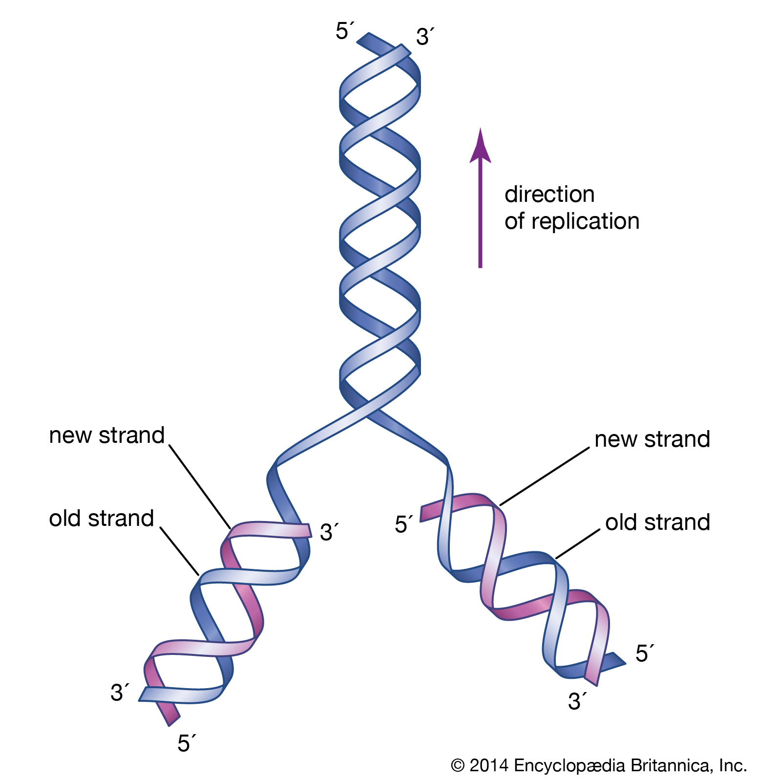
DNA replication is a semiconservative process in which the two strands are separated and new complementary strands are generated independently, resulting in two exact copies of the original DNA molecule. Each copy thus contains one strand that is derived from the parent and one newly synthesized strand. Replication begins at a specific point on a chromosome called an origin, proceeds in both directions along the strand, and ends at a precise point. In the case of circular chromosomes, the end is reached automatically when the two extending chains meet, at which point specific proteins join the strands. DNA polymerases cannot initiate replication at the end of a DNA strand; they can only extend preexisting oligonucleotide fragments called primers. Therefore, in linear chromosomes, special mechanisms initiate and terminate DNA synthesis to avoid loss of information. The initiation of DNA synthesis is usually preceded by synthesis of a short RNA primer by a specialized RNA polymerase called primase. Following DNA replication, the initiating primer RNAs are degraded.
The two DNA strands are replicated in different fashions dictated by the direction of the phosphodiester bond. The leading strand is replicated continuously by adding individual nucleotides to the 3′ end of the chain. The lagging strand is synthesized in a discontinuous manner by laying down short RNA primers and then filling the gaps by DNA polymerase, such that the bases are always added in the 5′ to 3′ direction. The short RNA fragments made during the copying of the lagging strand are degraded when no longer needed. The two newly synthesized DNA segments are joined by an enzyme called DNA ligase. In this way, replication can proceed in both directions, with two leading strands and two lagging strands proceeding outward from the origin.
Enzymes of replication
DNA polymerase adds single nucleotides to the 3′ end of either an RNA or a DNA molecule. In the prokaryote E. coli, there are three DNA polymerases; one is responsible for chromosome replication, and the other two are involved in the resynthesis of DNA during damage repair. DNA polymerases of eukaryotes are even more complicated. In human cells, for instance, more than five different DNA polymerases have been characterized. Separate polymerases catalyze the synthesis of the leading and lagging strands in human cells, and a separate polymerase is responsible for replication of mitochondrial DNA. The other polymerases are involved in the repair of DNA damage.
A number of other proteins are also essential for replication. Proteins called DNA helicases help to separate the two strands of DNA, and single-stranded DNA binding proteins stabilize them during opening prior to being copied. The opening of the DNA helix introduces considerable strain in the form of supercoiling, a movement that is subsequently relaxed by enzymes called topoisomerases (see above Supercoiling). A special RNA polymerase called primase synthesizes the primers needed at the origin to begin transcription, and DNA ligase seals the nicks formed between individual fragments.
The ends of linear eukaryotic chromosomes are marked by special sequences called telomeres that are synthesized by a special DNA polymerase called telomerase. This enzyme contains an RNA component that serves as a template for the exact sequence found at the ends of chromosomes. Multiple copies of a short sequence within the telomerase-associated RNA are made and added to the telomere ends. This has the effect of preventing shortening of the DNA chain that would otherwise occur during replication.
Single-stranded viral genomes, mitochondrial genomes, and some viral genomes are replicated in specialized ways. Several viruses such as adenovirus use a nucleotide covalently bound to a protein as a primer, and the protein remains covalently bound to the DNA after replication. Many single-stranded viruses use a rolling circle mechanism of replication whereby a double-stranded copy of the virus is first made. The replicating machinery then copies the nonviral strand in a continuous fashion, generating long single-stranded DNA from which full-length viral DNA strands are excised by specialized nucleases. |

|
|
|
|
Recombination
Recombination (B)
Recombination is the principal mechanism through which variation is introduced into populations. For example, during meiosis, the process that produces sex cells (sperm or eggs), homologous chromosomes—one derived from the mother and the equivalent from the father—become paired, and recombination, or crossing-over, takes place. The two DNA molecules are fragmented, and similar segments of the chromosome are shuffled to produce two new chromosomes, each being a mosaic of the originals. The pair separates so that each sperm or egg receives just one of the shuffled chromosomes. When sperm and egg fuse, the normal set of two copies of each chromosome is restored.
There are two forms of recombination, general and site-specific. General recombination typically involves cleavage and rejoining at identical or very similar sequences. In site-specific recombination, cleavage takes place at a specific site into which DNA is usually inserted. General recombination occurs among viruses during infection, in bacteria during conjugation, during transformation whereby DNA is directly introduced into cells, and during some types of repair processes. Site-specific recombination is frequently involved in the parasitic distribution of DNA segments throughout genomes. Many viruses, as well as special segments of DNA called transposons, rely on site-specific recombination to multiply and spread. The two processes are described in greater detail below.
General recombination
General recombination, also called homologous recombination, involves two DNA molecules that have long stretches of similar base sequences. The DNA molecules are nicked to produce single strands; these subsequently invade the other duplex, where base pairing leads to a four-stranded DNA structure. The cruciform junction within this structure is called a Holliday junction, named after Robin Holliday, who proposed the original model for homologous recombination in 1964. The Holliday junction travels along the DNA duplex by “unzipping” one strand and reforming the hydrogen bonds on the second strand. Following this branch migration, the two duplexes can be nicked again, allowing them to separate. Finally, the nicks are repaired by DNA ligase. The result is two DNA duplexes in which the segment between the two nicks has been replaced. The enzymes involved in recombination have been characterized best in the prokaryote E. coli. A key enzyme is RecA, which catalyzes the strand invasion process. RecA coats single-stranded DNA and facilitates its pairing with a double-stranded DNA molecule containing the same sequence, which produces a loop structure.
Another protein, known as RecBC, is important for the recombination process. Functioning at free ends of DNA, RecBC catalyzes an unwinding-rewinding reaction as it traverses the length of the molecule. Since unwinding is faster than rewinding, a loop is produced behind the enzyme that facilitates subsequent pairing with another DNA molecule. A number of other proteins are also important for recombination, including single-stranded DNA binding proteins that stabilize single-stranded DNA, DNA polymerase to repair any gaps that might be formed, and DNA ligase to reseal the nicks after recombination is complete. The details of eukaryotic recombination are expected to parallel those found in E. coli, although the highly compact chromatin structure in eukaryotes makes the process more complicated.
It is important to note that the initial product of recombination between two regions of DNA that are similar but not identical will be a “ heteroduplex”—that is, a molecule in which mismatched bases will be present at some positions in the helix. Thus, in the specialized recombination that takes place during meiosis, one round of replication is necessary before the mosaic chromosomes produced by recombination are properly matched. Enzymes are present in cells that specifically recognize and repair mismatches, so that the initial products of recombination can sometimes be repaired before they are replicated. In such cases the final products of replication will not be true reciprocal events, but rather one of the original parental molecules will appear to have been maintained to the exclusion of the other—a process called gene conversion.
Recombination also functions occasionally to repair lesions in DNA. If one chromosome of a pair becomes irreversibly damaged, the information from the other chromosome can be copied and inserted by recombination to provide a correct replacement of the damaged section. The key idea here is that sequences flanking the damage from a sister chromosome can base-pair with the corresponding sequences on the damaged chromosome, thus allowing replication to copy the correct sequence and repair the lesion.
Site-specific recombination
Site-specific recombination involves very short specific sequences that are recognized by proteins. Long DNA sequences such as viral genomes, drug-resistance elements, and regulatory sequences such as the mating type locus in yeast can be inserted, removed, or inverted, having profound regulatory effects. More than any other mechanism, site-specific recombination is responsible for reshaping genomes. For example, the genomes of many higher organisms, including plants and humans, show evidence that transposable elements have been constantly inserted throughout the genome and even into one another from time to time.
One example of site-specific recombination is the integration of DNA from bacteriophage λ into the chromosome of E. coli. In this reaction, bacteriophage λ DNA, which is a linear molecule in the normal phage, first forms a circle and then is cleaved by the enzyme λ-integrase at a specific site called the phage attachment site. A similar site on the bacterial chromosome is cut by integrase to give ends with the identical extension. Because of the complementarity between these two ends, they can be rejoined so that the original circular λ chromosome is inserted into the chromosome of the E. coli bacterium. Once integrated, the phage can be held in an inactive state until signals are generated that reverse the process, allowing the phage genome to escape and resume its normal life cycle of growth and spread into other bacteria. This site-specific recombination process requires only λ-integrase and one host DNA binding protein called the integration host factor. A third protein, called excisionase, recognizes the hybrid sites formed on integration and, in conjunction with integrase, catalyzes an excision process whereby the λ chromosome is removed from the bacterial chromosome.
A similar but more widespread version of DNA integration and excision is exhibited by the transposons, the so-called jumping genes. These elements range in size from fewer than 1,000 to as many as 40,000 base pairs. Transposons are able to move from one location in a genome to another, as first discovered in corn (maize) during the 1940s and ’50s by Barbara McClintock, whose work won her a Nobel Prize in 1983. Most, if not all, transposons encode an enzyme called transposase that acts much like λ-integrase by cleaving the ends of the transposon as well as its target site. Transposons differ from bacteriophage λ in that they do not have a separate existence outside of the chromosome but rather are always maintained in an integrated site. Two types of transposition can occur—one in which the element simply moves from one site in the chromosome to another and a second in which the transposon is replicated prior to moving. This second type of transposition leaves behind the original copy of the transposon and generates a second copy that is inserted elsewhere in the genome. Known as replicative transposition, this process is the mechanism responsible for the vast spread of transposable elements in many higher organisms.
The simplest kinds of transposons merely contain a copy of the transposase with no additional genes. They behave as parasitic elements and usually have no known associated function that is advantageous to the host. More often, transposable elements have additional genes associated with them—for example, antibiotic resistance factors. Antibiotic resistance typically occurs when an infecting bacterium acquires a plasmid that carries a gene encoding resistance to one or more antibiotics. Typically, these resistance genes are carried on transposable elements that have moved into plasmids and are easily transferred from one organism to another. Once a bacterium picks up such a gene, it enjoys a great selective advantage because it can grow in the presence of the antibiotic. Indiscriminate use of antibiotics actually promotes the buildup of these drug-resistant plasmids and strains. |

|
|
|
|
Repair
Repair (B)
It is extremely important that the integrity of DNA be maintained in order to ensure the accurate workings of a cell over its lifetime and to make certain that genetic information is accurately passed from one generation to the next. This maintenance is achieved by repair processes that constantly monitor the DNA for lesions and activate appropriate repair enzymes. As described in the section General recombination, serious lesions in DNA such as pyrimidine dimers or gaps can be repaired by recombination mechanisms, but there are many other repair mechanisms.
One important mechanism is that of mismatch repair, which has been studied extensively in E. coli. The system is directed by the presence of a methyl group within the sequence GATC on the template strand. Comparable systems for mismatch repair also operate in eukaryotes, though the template strand is not marked by methyl groups. In fact, lesions within the genes for human mismatch repair systems are known to be responsible for many cancers. Loss of the mismatch repair system allows mutations to build up quickly and eventually to affect the genes that cause cells to divide. As a result, cells divide in an uncontrolled manner and become cancerous.
Once replication is complete, the most common kind of damage to nucleic acids is one in which the normal A, C, G, and T bases are changed into chemically modified bases that usually differ significantly from their natural counterparts. The only exceptions are the deamination of cytosine to uracil and the deamination of 5-methylcytosine to thymine. In these cases the product is a G:U or G:T mismatch. Specific enzymes called DNA glycosylases can recognize uracil in DNA or the thymine in a G:T mismatch and can selectively remove the base by cleaving the bond between the base and the deoxyribose sugar. Many of these enzymes are specific for the different chemically modified bases that may be present in DNA.
Another common means of repairing DNA lesions is by an excision repair pathway. Enzymes recognize damage within DNA, probably by detecting an altered conformation of DNA, and then nick the strand on either side of the lesion, allowing a small single-stranded DNA to be excised. DNA polymerase and DNA ligase then repair the single-stranded gap. In all of these systems, the presence of an abnormal base signifies which strand is to be repaired, and the complementary strand is used as the template to ensure the accuracy of repair. |

|
|
|
|
RNA metabolism
RNA metabolism (B)
RNA provides the link between the genetic information encoded in DNA and the actual workings of the cell. Some RNA molecules such as the rRNAs and the snRNAs (described in the section Types of RNA) become part of complicated ribonucleoprotein structures with specialized roles in the cell. Others such as tRNAs play key roles in protein synthesis, while mRNAs direct the synthesis of proteins by the ribosome. Three distinct phases of RNA metabolism occur. First, selected segments of the genome are copied by transcription to produce the precursor RNAs. Second, these precursors are processed to become functionally mature RNAs ready for use. When these RNAs are mRNAs, they are then used for translation. Third, after use the RNAs are degraded, and the bases are recycled. Thus, transcription is the process where a specific segment of DNA, a gene, is copied into a specific RNA that encodes a single protein or plays a structural or catalytic role. Translation is the decoding of the information within mRNA molecules that takes place on a specialized structure called a ribosome. There are important differences in both transcription and translation between prokaryotic and eukaryotic organisms. |

|
|
|
|
Transcription
Transcription (B)
Small segments of DNA are transcribed into RNA by the enzyme RNA polymerase, which achieves this copying in a strictly controlled process. The first step is to recognize a specific sequence on DNA called a promoter that signifies the start of the gene. The two strands of DNA become separated at this point, and RNA polymerase begins copying from a specific point on one strand of the DNA using a ribonucleoside 5′-triphosphate to begin the growing chain. Additional ribonucleoside triphosphates are used as the substrate, and, by cleavage of their high-energy phosphate bond, ribonucleoside monophosphates are incorporated into the growing RNA chain. Each successive ribonucleotide is directed by the complementary base pairing rules of DNA. Thus, a C in DNA directs the incorporation of a G into RNA, G is copied into C, T into A, and A into U. Synthesis continues until a termination signal is reached, at which point the RNA polymerase drops off the DNA, and the RNA molecule is released. In some cases this RNA molecule is the final mRNA. In other cases it is a pre-mRNA and requires further processing before it is ready for translation by the ribosome. Ahead of many genes in prokaryotes, there are signals called “ operators” where specialized proteins called repressors bind to the DNA just upstream of the start point of transcription and prevent access to the DNA by RNA polymerase. These repressor proteins thus prevent transcription of the gene by physically blocking the action of the RNA polymerase. Typically, repressors are released from their blocking action when they receive signals from other molecules in the cell indicating that the gene needs to be expressed. Ahead of some prokaryotic genes are signals to which activator proteins bind that positively induce transcription.
Transcription in higher organisms is more complicated. First, the RNA polymerase of eukaryotes is a more complicated enzyme than the relatively simple five-subunit enzyme of prokaryotes. In addition, there are many more accessory factors that help to control the efficiency of the individual promoters. These accessory proteins are called transcription factors and typically respond to signals from within the cell that indicate whether transcription is required. In many human genes, several transcription factors may be needed before transcription can proceed efficiently. A transcription factor can cause either repression or activation of gene expression in eukaryotes.
During transcription, only one strand of the DNA is usually copied. This is called the template strand, and the RNA molecules produced are single-stranded. The DNA strand that would correspond to the mRNA is called the coding or sense strand, and it is not unusual for this to change from one gene to the next. In eukaryotes the initial product of transcription is called a pre-mRNA, which is extensively spliced before the mature mRNA is produced, ready for translation by the ribosome. |

|
|
|
|
Translation
Translation (B)
The process of translation uses the information present in the nucleotide sequence of mRNA to direct the synthesis of a specific protein for use by the cell. Translation takes place on the ribosomes—complex particles in the cell that contain RNA and protein. In prokaryotes the ribosomes are loaded onto the mRNA while transcription is still ongoing. Near the 5′ end of the mRNA, a short sequence of nucleotides signals the starting point for translation. It contains a few nucleotides called a ribosome binding site, or Shine-Dalgarno sequence. In E. coli the tetranucleotide GAGG is sufficient to serve as a binding site. This typically lies five to eight bases upstream of an initiation codon. The mRNA sequence is read three bases at a time from its 5′ end toward its 3′ end, and one amino acid is added to the growing chain from its respective aminoacyl tRNA, until the complete protein chain is assembled. Translation stops when the ribosome encounters a termination codon, normally UAG, UAA, or UGA. Special release factors associate with the ribosome in response to these codons, and the newly synthesized protein, tRNAs, and mRNA all dissociate. The ribosome then becomes available to interact with another mRNA molecule.
In eukaryotes the essence of protein synthesis is the same, although the ribosomes are more complicated. As with prokaryotic initiation, the signal sequence interacts with the 3′ end of the small subunit rRNA during formation of the initiation complex.
The issue of fidelity is important during protein synthesis, but it is not as crucial as fidelity during replication. One mRNA molecule can be translated repeatedly to give many copies of the protein. When an occasional protein is mistranslated, it usually does not fold properly and is then degraded by the cellular machinery. However, proofreading mechanisms exist within the ribosome to ensure accurate pairing between the codon in the mRNA and the anticodon in the tRNA.
One of the crowning achievements of molecular biology was the elucidation during the 1960s of the genetic code. Principals in this effort were Har G. Khorana and Marshall W. Nirenberg, who shared a Nobel Prize in 1968. Khorana and Nirenberg used artificial templates and protein synthesizing systems in the test tube to determine the coding potential of all 64 possible triplet codons (see the table). The key feature of the genetic code is that the 20 amino acids are encoded by 61 codons. Thus, there is degeneracy in the code such that one amino acid is often specified by more than one codon. In the case of serine and leucine, six codons can be used for each. Among organisms that have been examined in detail, the code appears to be almost universal, from bacteria through archaea to eukaryotes. The known exceptions are found in the mitochondria of humans and many other organisms as well as in some species of bacteria. The structure within the genetic code whereby many amino acids are uniquely coded by the first two bases of the codon strongly suggests that the code has itself evolved from a more primitive code involving 16 dinucleotides. How the individual amino acids became associated with the different codons remains a matter of speculation. |
| |
|

|
|
|
|
| |

|
|
|
|
|
|
Nucleic acid
Nucleic acid (W)
Nucleic acids are the biopolymers, or large biomolecules, essential to all known forms of life. The term nucleic acid is the overall name for DNA and RNA. They are composed of nucleotides, which are the monomers made of three components: a 5-carbon sugar, a phosphate group and a nitrogenous base. If the sugar is a compound ribose, the polymer is RNA (ribonucleic acid); if the sugar is derived from ribose as deoxyribose, the polymer is DNA (deoxyribonucleic acid).
Nucleic acids are the most important of all biomolecules. These are found in abundance in all living things, where they function to create and encode and then store information of every living cell of every life-form organism on Earth. In turn, they function to transmit and express that information inside and outside the cell nucleus — to the interior operations of the cell and ultimately to the next generation of each living organism. The encoded information is contained and conveyed via the nucleic acid sequence, which provides the 'ladder-step' ordering of nucleotides within the molecules of RNA and DNA.
Strings of nucleotides are bonded to form helical backbones — typically, one for RNA, two for DNA — and assembled into chains of base-pairs selected from the five primary, or canonical, nucleobases, which are: adenine, cytosine, guanine, thymine, and uracil. Thymine occurs only in DNA and uracil only in RNA. Using amino acids and the process known as protein synthesis, the specific sequencing in DNA of these nucleobase-pairs enables storing and transmitting coded instructions as genes. In RNA, base-pair sequencing provides for manufacturing new proteins that determine the frames and parts and most chemical processes of all life forms. |
|
| |
History
|
|
Naked nucleic acid
Naked nucleic acid (W)
Naked NA refers to NA that is not associated with proteins, lipids, or any other molecule to help "protect it". Naked DNA can be found when transcriptional bursting is occurring. |
|
|
|
Occurrence and nomenclature
Occurrence and nomenclature (W)
The term nucleic acid is the overall name for DNA and RNA, members of a family of biopolymers, and is synonymous with polynucleotide. Nucleic acids were named for their initial discovery within the nucleus, and for the presence of phosphate groups (related to phosphoric acid). Although first discovered within the nucleus of eukaryotic cells, nucleic acids are now known to be found in all life forms including within bacteria, archaea, mitochondria, chloroplasts, and viruses (There is debate as to whether viruses are living or non-living). All living cells contain both DNA and RNA (except some cells such as mature red blood cells), while viruses contain either DNA or RNA, but usually not both. The basic component of biological nucleic acids is the nucleotide, each of which contains a pentose sugar (ribose or deoxyribose), a phosphate group, and a nucleobase. Nucleic acids are also generated within the laboratory, through the use of enzymes (DNA and RNA polymerases) and by solid-phase chemical synthesis. The chemical methods also enable the generation of altered nucleic acids that are not found in nature, for example peptide nucleic acids. |

|
|
|
|
Molecular composition and size
Molecular composition and size (W)
Nucleic acids are generally very large molecules. Indeed, DNA molecules are probably the largest individual molecules known. Well-studied biological nucleic acid molecules range in size from 21 nucleotides (small interfering RNA) to large chromosomes (human chromosome 1 is a single molecule that contains 247 million base pairs).
In most cases, naturally occurring DNA molecules are double-stranded and RNA molecules are single-stranded. There are numerous exceptions, however — some viruses have genomes made of double-stranded RNA and other viruses have single-stranded DNA genomes, and, in some circumstances, nucleic acid structures with three or four strands can form.
Nucleic acids are linear polymers (chains) of nucleotides. Each nucleotide consists of three components: a purine or pyrimidine nucleobase (sometimes termed nitrogenous base or simply base), a pentose sugar, and a phosphate group.
The substructure consisting of a nucleobase plus sugar is termed a nucleoside. Nucleic acid types differ in the structure of the sugar in their nucleotides – DNA contains 2′-deoxyribose while RNA contains ribose (where the only difference is the presence of a hydroxyl group). Also, the nucleobases found in the two nucleic acid types are different: adenine, cytosine, and guanine are found in both RNA and DNA, while thymine occurs in DNA and uracil occurs in RNA.
The sugars and phosphates in nucleic acids are connected to each other in an alternating chain (sugar-phosphate backbone) through phosphodiester linkages. In conventional nomenclature, the carbons to which the phosphate groups attach are the 3′-end and the 5′-end carbons of the sugar. This gives nucleic acids directionality, and the ends of nucleic acid molecules are referred to as 5′-end and 3′-end. The nucleobases are joined to the sugars via an N-glycosidic linkage involving a nucleobase ring nitrogen (N-1 for pyrimidines and N-9 for purines) and the 1' carbon of the pentose sugar ring.
Non-standard nucleosides are also found in both RNA and DNA and usually arise from modification of the standard nucleosides within the DNA molecule or the primary (initial) RNA transcript. Transfer RNA (tRNA) molecules contain a particularly large number of modified nucleosides. |

|
|
|
|
Topology
Topology (W)
Double-stranded nucleic acids are made up of complementary sequences, in which extensive Watson-Crick base pairing results in a highly repeated and quite uniform double-helical three-dimensional structure. In contrast, single-stranded RNA and DNA molecules are not constrained to a regular double helix, and can adopt highly complex three-dimensional structures that are based on short stretches of intramolecular base-paired sequences including both Watson-Crick and noncanonical base pairs, and a wide range of complex tertiary interactions.
Nucleic acid molecules are usually unbranched and may occur as linear and circular molecules. For example, bacterial chromosomes, plasmids, mitochondrial DNA, and chloroplast DNA are usually circular double-stranded DNA molecules, while chromosomes of the eukaryotic nucleus are usually linear double-stranded DNA molecules. Most RNA molecules are linear, single-stranded molecules, but both circular and branched molecules can result from RNA splicing reactions. The total amount of pyrimidines is equal to the total amount of purines. The diameter of the helix is about 20Å. |

|
|
|
|
Sequences
Sequences (W)
One DNA or RNA molecule differs from another primarily in the sequence of nucleotides. Nucleotide sequences are of great importance in biology since they carry the ultimate instructions that encode all biological molecules, molecular assemblies, subcellular and cellular structures, organs, and organisms, and directly enable cognition, memory, and behavior (see Genetics). Enormous efforts have gone into the development of experimental methods to determine the nucleotide sequence of biological DNA and RNA molecules, and today hundreds of millions of nucleotides are sequenced daily at genome centers and smaller laboratories worldwide. In addition to maintaining the GenBank nucleic acid sequence database, the National Center for Biotechnology Information (NCBI, https://www.ncbi.nlm.nih.gov) provides analysis and retrieval resources for the data in GenBank and other biological data made available through the NCBI web site. |
|
|
|
| |
Types
|
Deoxyribonucleic acid
Deoxyribonucleic acid (W)
Deoxyribonucleic acid (DNA) is a nucleic acid containing the genetic instructions used in the development and functioning of all known living organisms. The DNA segments carrying this genetic information are called genes. Likewise, other DNA sequences have structural purposes or are involved in regulating the use of this genetic information. Along with RNA and proteins, DNA is one of the three major macromolecules that are essential for all known forms of life. DNA consists of two long polymers of simple units called nucleotides, with backbones made of sugars and phosphate groups joined by ester bonds. These two strands run in opposite directions to each other and are, therefore, anti-parallel.
Attached to each sugar is one of four types of molecules called nucleobases (informally, bases). It is the sequence of these four nucleobases along the backbone that encodes information. This information is read using the genetic code, which specifies the sequence of the amino acids within proteins. The code is read by copying stretches of DNA into the related nucleic acid RNA in a process called transcription. Within cells, DNA is organized into long structures called chromosomes. During cell division these chromosomes are duplicated in the process of DNA replication, providing each cell its own complete set of chromosomes. Eukaryotic organisms (animals, plants, fungi, and protists) store most of their DNA inside the cell nucleus and some of their DNA in organelles, such as mitochondria or chloroplasts. In contrast, prokaryotes (bacteria and archaea) store their DNA only in the cytoplasm. Within the chromosomes, chromatin proteins such as histones compact and organize DNA. These compact structures guide the interactions between DNA and other proteins, helping control which parts of the DNA are transcribed. |

|
|
|
|
Ribonucleic acid
Ribonucleic acid (W)
Ribonucleic acid (RNA) functions in converting genetic information from genes into the amino acid sequences of proteins. The three universal types of RNA include transfer RNA (tRNA), messenger RNA (mRNA), and ribosomal RNA (rRNA).
Messenger RNA acts to carry genetic sequence information between DNA and ribosomes, directing protein synthesis.
Ribosomal RNA is a major component of the ribosome, and catalyzes peptide bond formation.
Transfer RNA serves as the carrier molecule for amino acids to be used in protein synthesis, and is responsible for decoding the mRNA. In addition, many other classes of RNA are now known. |
|
|
|
|
| |
See also
|
|

|
|
|
|
|
|

